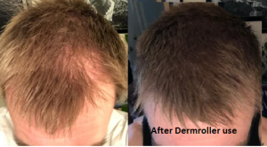
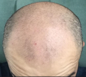
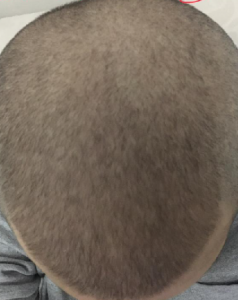
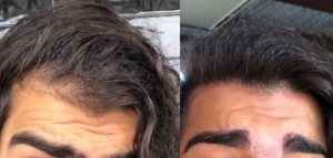
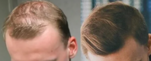
The side effect and complications of oral minoxidil are not so much of a problem if the dose is kept low. Too many doctors prescribe higher doses and patients get blood pressure problems, even some cardiac complications. See article here: https://www.jaad.org/article/S0190-9622(19)30685-1/fulltext
Development of Liposomal Systems of Finasteride for Topical Applications: Design, Characterization, and in Vitro Evaluation
- PMID: 18161632
- DOI: 10.1080/10837450701481181
Abstract
Finasteride (FNS) is a “drug of choice” for benign prostate hypertrophy and prostate cancer. The drug has also been reported to be useful orally in the treatment of some difficult-to-treat androgen-dependent skin disorders, such as seborrhea, acne, hirsutism, and androgenetic alopecia. However, the ideal route for its administration (i.e., topical) remains unexplored. This has logically suggested the search for strategic formulation approaches to make the drug effective on topical applications, hitherto unexplored. The present study targets the encasement of drug molecules in the interiors of vesicular compartments (liposomes) made up of hydrogenated phospholipids, as an attempt toward the development of a trans-epidermal therapeutic system of FNS. Multilamellar drug-loaded liposomes were prepared by thin-film hydration with sonication method and optimized with respect to drug payload, entrapment efficiency, and size by formulating different vesicular compositions under different process conditions. The vesicular systems consisting of saturated phospholipid (100 mg), cholesterol (50 mg), and FNS (5 mg) showed highest drug payload (2.9 mg/100 mg of total lipids), and drug entrapment efficiency (88.6%). Mean (+/-SD) vesicle size of the prepared liposomes was found to be 3.66+/-1.6 microm. Significantly higher skin permeation of FNS through excised abdominal mice skin of FNS was achieved from the liposomal formulations vis-à-vis corresponding solution and conventional gels. Liposomal FNS formulations also showed more than fivefold higher deposition of drug in skin than the corresponding plain drug solution and conventional gel. Stability studies indicated that the liposomal formulations were quite stable in the refrigerated conditions for 2 months with negligible drug leakage or vesicle size alteration during the storage period. Results of the current studies with FNS-loaded vesicular systems project the high plausibility of a topical liposomal formulation for effective localized delivery of Finasteride.
This is a compilation of information gleamed from a Reddit poster who put together a series of articles which may point to risks brought on by finasteride in pregnancy. I will quote the entire post and all of the references for you to draw your own conclusions. I have commented on two of the references. Many of the articles are controversial. We have known for some time, that finasteride can impact the fetus on sexual identification. Many hermaphrodites come from women who have ingested finasteride or any similar compound during the first trimester of pregnancy, so items #1-6 have to my knowledge been previously identified. But to develop such abnormalities women were required to have significant exposure to finasteride with some of it being absorbed in their body during pregnancy. In my own practice, I have seen hundreds of men father normal children while they were on finasteride, so there is no 1:1 relationship when a man is taking finasteride and abnormal children and have not seen any patient come to me complaining of an abnormal child while they conceived a baby while on the drug. Of course, that is just my experience and does not guarantee the safety of this drug. I leave that up to the FDA. The rest I will leave for you to judge after reading the material below.
Risks of Finasteride Exposure in Pregnancy

Finasteride, Propecia and Duasteride labels warn that pregnant women must not be exposed to the drug. This includes by taking it, touching a tablet, or by being exposed to semen from a man who is taking it.1
This is because if the active ingredient in Finasteride is absorbed by a woman who is pregnant with a male baby, it can cause the male baby to be born with abnormalities of the sex organs. Finasteride blocks DHT, which is necessary for male baby organs to form and grow. Prenatal Finasteride exposure has also been shown to affect cognitive function in both male and female babies.
What are the specific problems? According to the research they include:
Higher rates of miscarriage
A small penis
Hypospadias
A small underdeveloped scrotum
A prominent midline raphe (scrotum doesn’t join normally)
Ectopic testicles (which don’t reach the scrotum)
Cognitive problems in both males AND females
Memory problems in both males AND females
Three sources detailing these effects are provided below.
Source 1: S. Prahalada (1997) Effects of Finasteride, a Type 2 5-Alpha Reductase Inhibitor, on Fetal Development in the Rhesus Monkey2
In this study pregnant female monkeys were orally given 2mg/kg/day. Every single one of their male babies had genital birth defects.
“The male external genital anomalies were characterised by hypospadias which were confined to the glans penis (5/6; 83.3%), preputial adhesion to the glans (6/6; 100%), a small underdeveloped scrotum (6/6; 100%), a small penis (5/6; 100%) and a prominent midline raphe (6/6; 100%).”
“An apparent incidence in the fetal loss rate was observed in the 2mg/kg/day (23.1%) and the 800ng/kg/day (20%) finasteride treated groups when compared to concurrent controls…”
Source 2: Christopher Bowman, et al (2003). Effects of in Utero Exposure to Finasteride on Androgen-Dependent Reproductive Development in the Male Rat3
In this study Finasteride is shown to impact testicular descent: “Prenatal finasteride exposure significantly impaired testicular descent. Approximately 3, 23, and 73% of the adult males displayed ectopic testes in the 1.0, 10, and 100 mg/kg/day dose groups, respectively… The data from the current study suggest that the conversion of T to DHT in the developing gubernaculum is necessary for normal testicular descent.”
Even in the smallest dose of 0.01 mg/kg/day in the last stages of gestation there were genital issues affecting anogenital distance (AGD) of the males after the rats were born. “Late gestational exposure to finasteride significantly decreased AGD of male offspring in a dose-responsive manner… the AGD of male offspring displayed significant decreases of 8, 16, 23, 25, and 33% in the 0.01, 0.1, 1.0, 10, and 100 mg/kg/day dose groups, respectively.”
“The dose-response curves for finasteride-induced malformations fell into two groups. External structural changes such as decreased AGD and increased nipple retention had similar dose-response curves. These external changes were almost linear over the entire dose range (0.01 to 100 mg/kg/day) and approached 100% incidence by the highest dose.”
The authors note that “The lack of a no observed effect level (NOEL) in the current study was consistent” with the study by Clark (1990) which showed effects at doses down to 0.003 mg/kg/day. (DR RASSMAN’S COMMENT: THIS STUDY WAS DONE IN MONKEYS ADMINISTERED FINASTERIDE IN VARIOUS DOSES DURING PREGNANCY. THIS IS NOT THE SAME AS THE MALE MONKEY BEING ON FINASTERIDE WHEN THE FEMALE MONKEY BECAME PREGNANT)
Source 3: Jason Paris et al (2012) Inhibition of 5?-reductase activity in late pregnancy decreases gestational length and fecundity and impairs object memory and central progestogen milieu of juvenile rat offspring4
“Finasteride significantly reduced the length of gestation and the number of pups per litter…”
“Prenatal finasteride treatment significantly reduced object recognition, decreased hippocampal 3?,5?-THP content… inhibiting the formation of 5?-reduced steroids during late gestation in rats reduces gestational length, the number of viable pups per litter, and impairs cognitive and neuroendocrine function in the juvenile offspring.” (THIS STUDY WAS DONE IN RATS, AGAIN IN PREGNANCY AND DOES NOT CORRESPOND TO A MALE RAT TAKING FINASTERIDE WHEN THE FEMALE RAT BECAME PREGNANT)
While some of these research exposures were a lot higher than would be expected from semen exposure, don’t risk exposing a baby to any amount of this extremely dangerous US FDA pregnancy category X drug.5 Studies have demonstrated fetal abnormalities and there is positive evidence of human fetal risk.
Links:
2.https://www.docme.su/doc/1721148/effects-of-finasteride–a-type-2-5-alpha-reductase-inhibi…
3.https://academic.oup.com/toxsci/article/74/2/393/1716348
4.https://www.ncbi.nlm.nih.gov/pmc/articles/PMC3196810/#__ffn_sectitle
Back in the 80’s & early 90’s this was an acceptable hairstyle to rock for men with hair loss. The shaved head has only been a more recent trend among balding dudes. At some point will the horseshoe be considered a more stylish option than shaving your head? I’m curious because I’m sick of balding dudes all having to rock the same haircut.
Balding with a shaved head works for many men. Some men get Scalp Micropigmentation (https://scalpmicropigmentation.com/ and look at the men on the front page. Also drill down on the bold men in the gallery). Either way, it is more what is in your head than what is on top of it.
I know this is going to sound weird, but my hairloss really started only 1 or 2 weeks after I began obsessively taking pictures of my temples, crown, etc and becoming anxious over it. Back then I seriously panicked thinking I had MPB as a teen, leading me to this sub. In retrospect, it looked perfectly fine. It then suddenly became much thinner, and since then my hairline, primarily the temples, has been steadily receding and my hair turned thinner, like a self-fulfilling prophecy.
MPB is certainly genetic, but what factors (like stress) could trigger it to start occurring earlier?
Some men practice denial, and one day they look and there it is! Are you really balding? See a good doctor and find out.
I have performed hair transplants on many men who will not take drugs of any type. The nice thing about a hair transplant, is that other than its progressive nature, there is no maintenance required for the transplanted hair to grow for your lifetime in 95% of men who get it.
Always look up the doctor and see his credentials (here are mine: https://newhair.com/doctors/rassman/ ). Ask to see his patients and meet them one-on-one because what you see is what you are going to get. It is not very complicated but too many doctors are ‘sold’ by salesmen who know how to convert you to get your money.
I weighed around 85-86kg before this, and now, two weeks and two days later from the initial depression episode starting, ended two days ago, I weigh around 80-81kg apparently according to the scales. I am concerned, thats alot of weight to lose in such a short time.
It is different with different people, but 15 pounds per months could trigger hair loss with the genes for balding on board. When the genes kick in, it rarely reverses.
There is no doubt that personalities change with balding, and when I do a hair transplant, they often change for the better radically. I see that in many celebrities I have done. They feel that the hair transplant not only changed their appearance but changed their effectiveness in their acting careers.
I’m sitting at about a Norwood 1.25 or so right now, and was wondering if I could restore it to at least a 1 reliably with only min since I caught it early. I’m also horrified of the dreaded shed making it appear worse, will this be lessened since I caught it early?
The younger you are, the better the chance that finasteride might work on the hairline. This does not mean restoring the juvenile hairline and reversing the mature hairline, but I am referring to real hairline recession in men under 22 years old. Although, I have seen this happen in older men under 30 less frequently.
No, these drugs kick in quickly but it may take months to see the value of their use.
Dr. Rassman’s comment: Dr. Cotsarelis is one of the most well known regenerative medicine experts in the world so this summary should has great interest for the readers of this blog. I congratulate Dr. Cotsarelis and Dr. Wang for this very interesting research project.
Source: News & views
Hair-bearing skin grown in a dish, Authors: Leo L. Wang & George Cotsarelis
Undifferentiated human stem cells have been coaxed to develop into skin-like structures in vitro. When engrafted onto mice, the structures produce hair — highlighting the potential
of the approach for regenerative therapies
When hair follicles were first generated from stem cells that had been isolated from adult mouse skin1 , Jay Leno — a former host of US talk show The Tonight Show — joked that scientists “cured baldness … at least in mice”. Sixteen years on, the current host will have the opportunity to mention that scientists have ‘cured’ baldness in humans, now that Lee et al. 2 , writing in Nature, have regenerated hair follicles from human stem cells. This achievement places us closer to generating a limitless supply of hair follicles that can be transplanted to the scalps of people who have thinning or no hair. Moreover, if the approach reaches the clinic, individuals who have wounds, scars and genetic skin diseases will have access to revolutionary treatments. Research into skin-tissue engineering began in 1975, when a landmark study showed that cells called keratinocytes could be isolated from the surface layer of a person’s skin (the epidermis)3 , and the cell population expanded in culture. Almost a decade later, sheets of keratinocytes isolated from people with burns were transplanted back to the individuals they came from as life-saving, permanent engraftments4 . This work was the foundation for another remarkable achievement in 2017, when a boy who had a genetic disease called junctional epidermolysis bullosa (which causes severe fragility of the skin) had his epidermis replaced with genetically corrected cells5 . For this type of cell-based approach to advance further, grafted skin must include more of the components found in normal skin: hair follicles, pigment-producing melanocyte cells, sweat glands, nerves, muscle, fat and immune cells, in addition to epidermal cells. Enter Lee and colleagues. The authors leveraged research from the fields of stemcell biology and hair-follicle development6 to generate near-complete skin organoids — self-organizing tissues grown in the laboratory that mimic developing skin. Organoids have been grown to imitate various organs, including the gut, lung, kidney and brain7 . Because organs consist of many cell types, organoids are typically formed from pluripotent stem cells, which have the ability to form all adult cell types. These can be embryonic stem cells or induced pluripotent stem cells, which are created by reprogramming adult cells to an embryonic-like state8 . The epidermis and the dermis — the skin’s other main component — are derived from different cell types in the early embryo. Lee et al. optimized the culture conditions needed to generate skin organoids containing both components from human pluripotent stem cells. The authors sequentially added growth factors to the stem cells. First, they used BMP4 and an inhibitor of the transcription factor TGF-? to induce formation of the epidermis. Next, they treated the cells with the growth factor FGF2 and an inhibitor of BMP, to induce the formation of cranial neural crest cells, which give rise to the dermis. The cells grew in a sphere. After more than 70 days, follicles began to appear, which ultimately produced hair (Fig. 1). Most hairs were pigmented by melanocytes, which also develop from the cranial neural crest. Tissues associated with hair follicles — such as sebaceous glands, nerves and their receptors, muscles and fat — developed, too, leading to the formation of remarkably complete skin in a dish9 . One missing component, however, was immune cells, which normally reside in and around hair follicles, and have diverse roles in the skin10. Lee and colleagues found that their organoids expressed genes that were characteristic of skin from the chin, cheek and ear. Interestingly, dermal cells on the scalp might also derive from the neural crest11. This suggests that the organoids might actually mimic scalp skin, and also highlights that it could be possible to tailor the authors’ protocol to generate skin that has the characteristics of Regenerative medicine Hair-bearing skin grown in a dish Leo L. Wang & George Cotsarelis Undifferentiated human stem cells have been coaxed to develop into skin-like structures in vitro. When engrafted onto mice, the structures produce hair — highlighting the potential of the approach for regenerative therapies. Pluripotent stem cells Epidermal layer Dermal layer Fat-cell layer Organoid Latent period Transplant Epidermal differentiation using BMP4 and TGF-? inhibitor Dermal differentiation using FGF2 and BMP4 inhibitor Baldness Wound healing Genetic skin disorders Hair follicle Figure 1 | Skin grown in vitro as a future clinical therapy. Lee et al. 2 grew human pluripotent stem cells (which can give rise to all cell types) into spherical skin-like structures called organoids in vitro. To achieve this, they treated the cells with growth factors (BMP4 and a TGF-? inhibitor) that promote growth of the skin’s epidermal layer and then with other growth factors (FGF2 and a BMP4 inhibitor) that induce formation of the dermal layer (a fat-cell layer also forms at this stage). After a long latent period (more than 70 days), the full complement of skin cells formed in the organoid, including around 50 hair follicles. When the organoids are implanted into skin, the hair follicles naturally orient themselves in the correct direction. It is possible that these organoids could be used to treat baldness and genetic skin disorders, and to promote wound healing. Nature | 1 News & views https://doi.org/10.1038/d41586-020-01568-2 © 2 0 2 0 S p ri n g e r N a t u r e Li mi t e d. Al l ri g h t s r e s e r v e d. different body sites, by altering the culture conditions in which the cells are grown. The group’s organoids will be a perfect tool for analyzing the roles of various biological pathways in skin development — small-molecule inhibitors or inhibitory RNA molecules can be used to block proteins or pathways and to investigate the effects on skin growth. The organoids can be used in combination with genome-wide association studies or other genetic data to analyze how particular genetic mutations alter skin development. They can also help to model diseases of the skin and hair and to screen experimental drugs for any toxicities and for their efficacy. Beyond these in vitro benefits, the authors demonstrated that the organoids have therapeutic potential in vivo. They transplanted the organoids onto immunodeficient mice (to ensure the graft was not rejected by the animals’ immune system), and showed that just over half of organoids go on to form hair, which is distributed over the surface of the graft. This illustrates the exciting potential of introducing skin organoids into wounds to encourage healing and prevent scarring, or transplanting them into areas lacking hair. However, several questions remain before this therapeutic approach becomes a reality. For instance, how efficiently and reproducibly do hairs develop? How many cells are needed to eventually form a hair follicle once grafted? Lee et al. began to answer the first of these questions by showing that a separate laboratory could grow hair in organoids using the same culture conditions. However, dealing with variability between individual stem cells and between the stem cells from different people are daunting challenges. The prolonged length of time required for organoids to develop hair follicles mimics fetal skin development12. Similarly, in both settings, the skin undergoes a latent ‘resting’ phase before follicles begin to grow. This is a fascinating area for future study. However, it took 140 days before organoids were ready for engraftment, which could impede the therapeutic potential of the work — someone with burns, for instance, cannot wait that long for a skin graft. Further studies to understand the molecular events taking place during this latent phase should provide strategies for accelerating this process using molecules that alter relevant signaling pathways. Several other aspects of the authors’ approach will also need to be optimized before it can move to the clinic. The hairs that grew in the current study were small; in future, further optimization of culture conditions will be needed to form large scalp hairs. Better characterization of some components used in the culture cocktail — such as a protein mixture called Matrigel — will be necessary to ensure that they comply with good manufacturing practices. And future work might need to move away from using pluripotent stem cells, which can have undesirable side effects, such as promoting tumor formation. An appealing alternative might be to use adult stem cells. Despite these caveats, Lee and colleagues’ study is a major step towards a ‘cure’ for baldness in humans, and paves a way towards other, greater therapeutic possibilities. At a minimum, it is worth a shout-out on a late-night show. The work holds great promise of clinical translation — we are confident that research will eventually see this promise realized. Leo L. Wang and George Cotsarelis are in the Department of Dermatology, University of Pennsylvania, Philadelphia, Pennsylvania 19104, USA. e-mails: leo.wang@pennmedicine.upenn.edu; cotsarel@pennmedicine.upenn.edu 1. Morris, R. J. et al. Nature Biotechnol. 22, 411–417 (2004). 2. Lee, J. et al. Nature https://doi.org/10.1038/s41586-020- 2352-3 (2020). 3. Rheinwald, J. G. & Green, H. Cell 6, 331–343 (1975). 4. Gallico, G. G. III, O’Connor, N. E., Compton, C. C., Kehinde, O. & Green, H. N. Engl. J. Med. 311, 448–451 (1984). 5. Hirsch, T. et al. Nature 551, 327–332 (2017). 6. Saxena, N., Mok, K.-W. & Rendl, M. Exp. Dermatol. 28, 332–344 (2019). 7. Rossi, G., Manfrin, A. & Lutolf, M. P. Nature Rev. Genet. 19, 671–687 (2018). 8. Takahashi, K. & Yamanaka, S. Cell 126, 663–676 (2006). 9. Plikus, M. V. et al. Science 355, 748–752 (2017). 10. Kobayashi, T. et al. Cell 176, 982–997 (2019). 11. Ziller, C. in Hair Research for the Next Millenium: Proc. 1st Tricont. Meet. Hair Res. Socs (eds Randall, V. A. et al.) 19–23 (Elsevier, 1996). 12. Pinkus, H. in The Biology of Hair Growth (eds Montagna, W. & Ellis, R. A.) 1–32 (Academic, 1958)