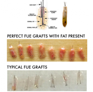
This photo contains Follicular Units that were excised from the donor area using two different techniques. The graphic at the top identifies all of the elements of a single Follicular Unit. This particular follicular unit has two hairs, one is what I am calling a ‘telogen’ hair (shorter hair below the skin is either starting to grow or shrinking down (more likely). On the rows of Follicular Units (grafts), you can count the number of hairs in each graft. On row two, the grafts were removed using a punch with subdermal tumescence. Note here in row two, that the hairs are surrounded by fat, especially prominent at the bulbs on the bottom of the grafts. Row three shows Follicular Units excised with a different punch. These grafts do not have much fat at the bottom and the three follicular units on the right have no fat at the bottom at all. Because there is no fat to protect them from drying once they are removed, they can not be exposed to the air for more than a few seconds without a risk drying and death. If these three grafts are placed with forceps, they might likely be damaged as the operator will have to use the forceps to squeeze the grafts at the bulb, possibly damaging the grafts. Placement with implanters, solves this problem.
